Drosophila third instar larval fillet labeled with AlexaFluor™647 for post-synaptic sites, AlexaFluor™555 conjugated to phalloidin, and AlexaFluor™488 labeling a subset of motor neurons. Sample courtesy Dr. Amicia Elliott, NIH/NIMH, Bethesda, MD (USA).
MIN6 cells grown as pseudoislets (pancreatic beta cells). DAPI (blue), Insulin (Alexa488, green), membrane receptor (Alexa594, red), phalloidin (Alexa647, white). Sample courtesy Dr. Rémy Bonnavion, MPI for Heart and Lung Research, Bad Nauheim (Germany).
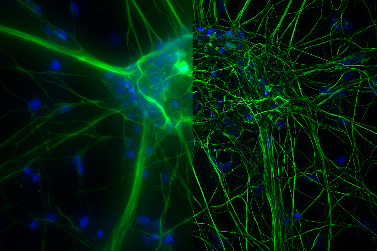
Cultured Cortical Neurons. Green, beta-III-tubulin; blue, Nuclei. Image stack of 59 planes for a volume of 21 µm. Sample Courtesy: FAN GmbH, Magdeburg (Germany).
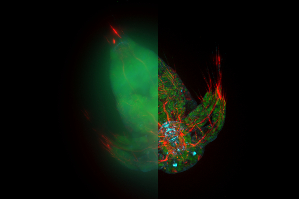
An organoid approximately 150 µm in diameter mounted onto a depression slide.
In this E12-14 mouse (wt sample), neurofilaments are stained in red to assess neuronal outgrowth. The mouse was cleared with the ScaleS reagent. Sample courtesy Yves Lutz, Centre d’imagerie, IGBMC (France).
Image of a locust ganglion, showing a maximum projection. Sample thickness: 110µm, data volume: 376 MB.
YFP mouse brain slices stained with GFAP-A647. Imaged with a THUNDER Imager Tissue. Courtesy Dr. Hong Xu, University of Pennsylvania, Philadelphia (USA).
Mouse kidney section with Alexa Fluor™ 488 WGA, Alexa Fluor™ 568 Phalloidin, and DAPI. Sample: FluoCells™ Prepared Slide #3, Thermo Fisher Scientific, Waltham, MA USA.
Zebrafish larvae (72 hours post fertilization). The blood vessels are indicated with green (fluorescence). Sample courtesy of Dr. Almary Guerra and Dr. Didier Stainier, Max Planck Institute for Heart and Lung Research, Bad Nauheim, Germany.
Mouse kidney section with Alexa Fluor™ 488 WGA, Alexa Fluor™ 568 Phalloidin, and DAPI. Sample: FluoCells™ Prepared Slide #3, Thermo Fisher Scientific, Waltham, MA USA.
Cultured VERO cells stained with STAR488 Vimentin (green), STAR580 Tom20 (yellow), and DAPI (blue). Sample courtesy of Abberior GmbH, Göttingen, Germany.
Smooth muscle around blood vessels in a 300 µm section of mouse lung. 1x objective. Courtesy of Dr. Mario Boehm, University of Giessen, Germany.
Zebrafish heart, DAPI (nuclei, blue), Tropomyosin (cardiomyocytes, red) and GFP (primordial cardiac layer, green). Courtesy of Anna Jazwinska, University of Fribourg, France.